The Relationship Between Procedural Memory and Purkinje Cells
A new study shows that a protein, known as SHISA6, plays an essential role in the formation of procedural memory for vestibulo-ocular reflex and eye movement conditioning.
Author: Abigail Bondurant
Neurophysiology
Background
Knowing how to do things, for example riding a bike, is a skill that is retrieved effortlessly and without conscious thought. The ‘knowing how’ to do this skill and others like it is because of procedural knowledge, or the consolidation of a memory through what is known as procedural memory (Perera, 2021). Procedural memory is a type of long-term memory that begins to form in the early stages of life. This process starts as soon as you begin to learn how to do things, such as crawling, walking, talking, and even eating, as an infant (Cherry, 2020). Procedural memories are formed and strengthened through the repeated use of connections that were first established between cells, known as neurons, in your brain (Cherry, 2020). The frequency of a learned action, like walking, sends signals between neurons that forms a connection known as a synaptic connection. Those synaptic connections become stronger until walking (the action) no longer requires conscious thought to complete (Cherry, 2020).
The area of the brain known as the cerebellum plays an important role in procedural memory because it controls balance, and it is used during the timing and coordination of skilled movements (Zimmermann, 2014; Guy-Evans, 2021). It also helps to establish the unconscious process during procedural learning (Perera, 2021). The main responsibility of the cerebellum is to control and regulate motor behaviors as well as coordinate respiratory rhythm, without conscious awareness (Guy-Evans, 2021; Encyclopedia Britannica, 2020). The dominant neurons within the cerebellum are Purkinje cells. Purkinje cells are large branching neurons and are essential for motor movement control (Encyclopedia Britannica, 2015). Purkinje cells continuously undergo long-term potentiation and long-term depression throughout life by either strengthening or weakening synaptic connections, respectively. These processes contribute coordination of motor movements and help to fine tune them (Paul & Limaiem, 2021).
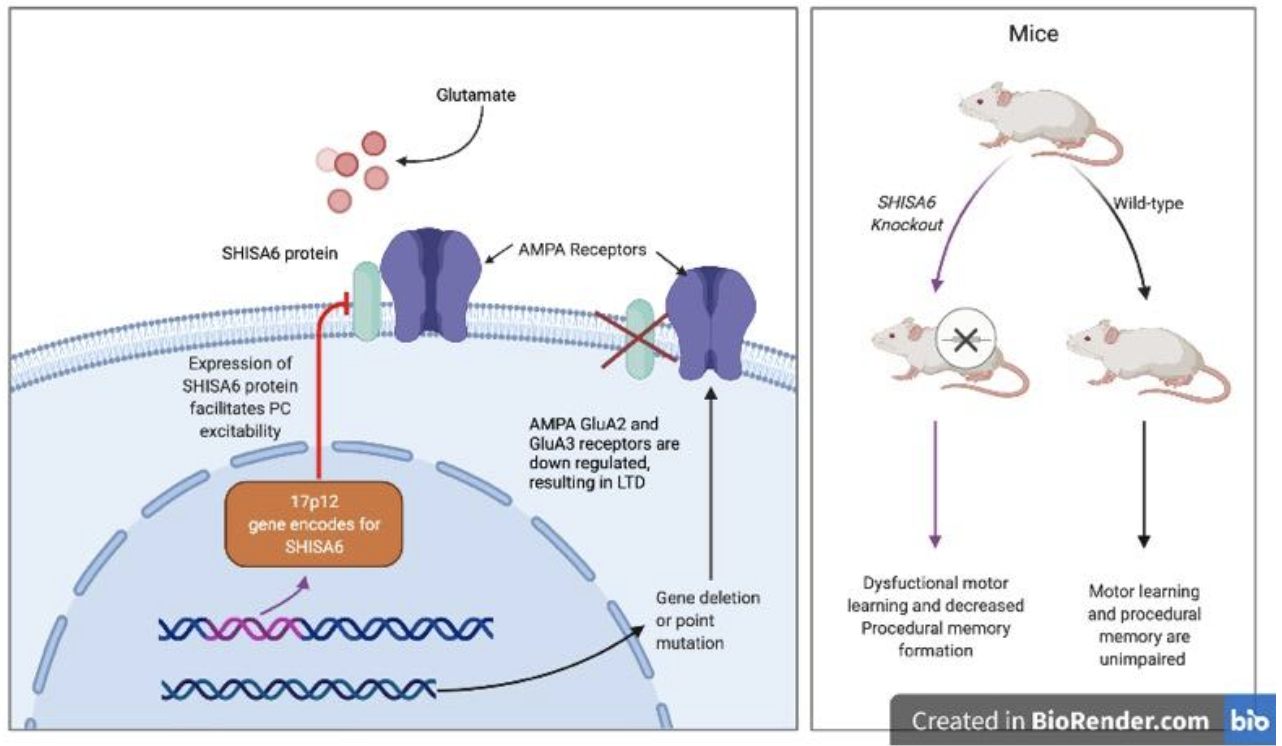
To further explore how the cerebellum influences the formation of procedural memories, a neuroscientist, Saša Peter, and a team of scientists used a mouse model that was genetically altered to assess how the expression of a protein, SHISA6, in Purkinje cells and their associated AMPA receptors, contributes to procedural memory formation.
Methodology
Peter et al focused on the vestibulo-ocular reflex and eye conditioning movements during their investigation into the relationship between the cerebellum and procedural memory. The vestibulo-ocular reflex is the ability to stabilize our perception of the world and remain balanced while our heads and eyes move continuously during everyday tasks. The vestibular system within the ear is responsible for the reflex, and information from this system is processed in the cerebellum (as well as other places) to aid in regulation of the reflex, coordination, and maintaining balance (Somisetty & Das, 2021).
Peter et al used two types of mice, a wild-type mouse and a SHISA6 knockout mouse and subjected each group to six visuo-vestibular mismatched training sessions in total. The goal was to increase vestibulo-ocular gain in the dark. The sessions consisted of a visual stimulus and vestibular stimulus each rotating in opposite directions and the eye movements of the mice adapting to the rotating stimuli was assessed. The wild-type mice were shown to have adapted fairly quickly due to an increase in their vestibulo-ocular reflex. The SHISA6 knockout mice were not able to adapt because of a lack of increase in their vestibulo-ocular reflex. A second training session was completed to assess if both groups of mice were able to reverse the direction their vestibulo-ocular reflex had adapted to. In this session, both the visual and vestibular stimulus rotated in the same direction. The SHISA6 knockout mice failed to demonstrate an ability to reverse their vestibulo-ocular phase, whereas the control mice were successful in this task.
The eye blinking of the mice was assessed using a conditioned response (light pulse) and an unconditioned response (corneal air puff). The SHISA6 knockout mice had a lower response (eye blinking) to both stimuli. The motor coordination of both groups was also assessed by evaluating the walking patterns of the mice on pressure sensitive rungs, and the SHISA6 knockout mice had a greater number of missteps which indicated an impairment.
Results
The study demonstrates the importance of the SHISA6 as an auxiliary protein for AMPA receptors in Purkinje cells. The results of the two training sessions and the eye blinking assessment indicates that SHISA6 protein is essential for procedural memory formation, which in this case motor coordination, maintaining balance, and coordinating eye movements and motor movements. The SHISA6 knockout mice were unable to orient themselves through coordination of eye and motor movements during either training sessions, and also showed impaired motor function. Because the protein, which is an auxiliary protein for AMPA receptors, wasn’t present in the knockout mice, the receptors were downregulated and there was a reduction in excitatory postsynaptic transmissions between other neurons and Purkinje cells.
Despite the fact there is very little known about the family of Shisa proteins, the interaction between SHISA6 and AMPA receptors has been demonstrated to contribute to the induction of long-term potentiation during procedural motor learning. The absence of the protein alters the function of Purkinje cells and impairs procedural memory that is essential for motor function and coordination. The study aimed to use a system, in this case vestibular system, that is responsible for maintaining balance and coordinating motor function during movements to show how the activity within Purkinje cells influences the procedural learning of said functions. The study provides support for the argument that SHISA6 proteins are essential for procedural memory formation because without them, there is decrease in excitatory activity and a downregulation of receptors that results in long-term depression.
[+] References
Britannica, T. Editors of Encyclopedia. (2015). Purkinje Cell. Encyclopedia Britannica.
https://www.britannica.com/science/Purkinje-cell.
Britannica, T. Editors of Encyclopedia. (2020). Hindbrain. Encyclopedia Britannica.
https://www.britannica.com/science/hindbrain
Cherry, K. (2020). How Procedural Memory Works. Verywell Mind.
https://www.verywellmind.com/what-is-procedural-memory-2795478
Guy-Evans, O. (2021). Cerebellum Functions, Structure, and Location. Simply Psychology.
www.simplypsychology.org/what-is-the-cerebellum.html
Paul, M.S. & Limaiem, F. (2021). Histology, Purkinje Cells. StatPearls [Internet]. StatPearls
Publishing. https://www.ncbi.nlm.nih.gov/books/NBK545154/
Perera, A. (2021). Procedural Memory. Simply Psychology.
Peter, S. et al. (2020). AMPAR Auxiliary Protein SHISA6 Facilitates Purkinje Cell Synaptic
Excitability and Procedural Memory Formation. Cell Reports, 31(2).
Somisetty, S. & Das, J.M. (2021). Neuroanatomy, Vestibulo-ocular Reflex. StatPearls
[Internet]. StatPearls Publishing. https://www.ncbi.nlm.nih.gov/books/NBK545297/
Zimmermann, K.A. (2014). Procedural Memory: Definition and Examples. LiveScience, Purch.
https://www.livescience.com/43595-procedural-memory-html.