Brain Cells Die So You Can See
This study shows that a specific type of brain cells die during vision development so people can have properly functioning binocular vision.
Author: Teyline McLean
Neurophysiology
Introduction
In a paper recently published in Neuron, Wang et al looked at the role that chandelier cells play in the development of binocular vision in mice. Chandelier cells can be hard to study and not much is known about their specific functions. During vision development, it is known that after eye opening, changes in neuronal activity can occur, but less is known about how vision develops before eye opening. This study found that the death and migration of chandelier cells in the visual cortex is mediated by retinal and neuronal activity and is an essential element of vision development before eye opening (Wang, 2021).
Background
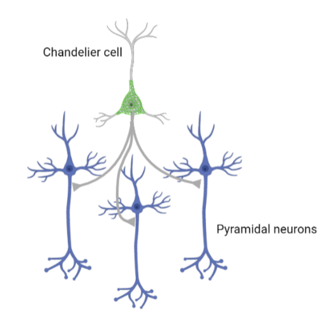
Chandelier cells, sometimes called axo-axonic cells, are GABAergic cortical interneurons. They were first discovered independently by Szentagothai and Jones in 1975. They are rare and hard to study as common neuronal markers also tend to label basket cells, making it hard to differentiate them (Woodruff, 2010). One chandelier cell can innervate hundreds of pyramidal neurons and one pyramidal cell can be connected to several chandelier cells. A very simplified schematic of this connection can be seen in Figure 1. Chandelier cells connect to the pyramidal neuron’s axon initial segment, where action potentials are generated (Lu, 2017). Pyramidal cells are glutamatergic, producing excitatory effects, and traverse multiple cortical layers and brain regions (Woodruff, 2010). Chandelier cells are commonly thought to produce powerful inhibitory effects, although there is some debate as to whether chandelier cells inhibit or excite pyramidal cells. There have been a few studies that used caged GABA to measure reversal potentials (EGABA) and found that chandelier cells can depolarize and cause glutamatergic excitatory postsynaptic potentials (EPSPs) in the pyramidal neurons after the activation (Szabadics, 2006), (Khirug, 2008), (Woodruff, 2010). This does not negate chandelier cell’s inhibition, but rather indicates that they can do both, producing a wider range of regulatory effects on pyramidal cells. It has also been reported that the location of the cells determines their primary effects, chandelier cells in cortical layer 2/3 depolarize when activated with GABA and in chandelier cells in CA1 hyperpolarize (Woodruff, 2009), (Woodruff, 2010).
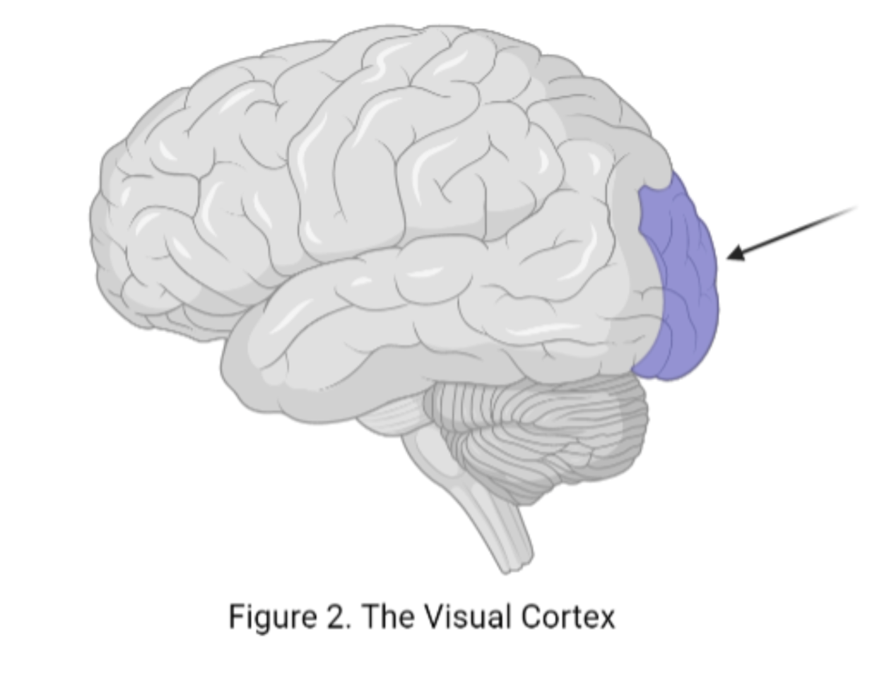
The visual cortex, located at the back of the brain, shown in Figure 2., is laid out into a primary area (V1) and lateral secondary visual areas (V2L). The binocular zone (BZ) is where these two areas intersect, and their neurons cross over into both areas (Wang 2021). Most people have two eyeballs that detect two different images of the world. Our brain combines these into the one cohesive image we see, this is binocular vision. Binocular vision is important for the accurate perception of the world around us, especially depth perception. It helps us determine how far away things are from us and this helps us do things like walk and pick up objects (Patla, 2002).
Methods
Wang et al fluorescently tagged chandelier cells in mice and looked at the amount and distribution of chandelier cells throughout the mice’s lifespan. They used several methods to manipulate the retinal and neural activity during development to observe the effects on chandelier cells and vision in mice. This was done by adding potassium (K+) channels, which increases the refractory period so action potentials cannot fire as often; adding an inhibitory DREADD receptor activated by CNO; administering tetrodotoxin (TTX), which blocks sodium (Na+) channels; and removing an eyeball, which eliminates the incoming visual stimulation to the brain. The mice’s vision was tested when chandelier cell development was disrupted to determine how much they could see (Wang 2021).
Results
Early in life, chandelier cells are evenly spread throughout the primary visual cortex in the V1, BZ, and V2L areas. As the mice age, chandelier cells are eliminated from the BZ, as they migrate out of the BZ and into the V1 and V2L regions. When retinal and neuronal activity is inhibited through any of the methods mentioned above, the chandelier cells are not eliminated and stay in the BZ, indicating that retinal and neuronal activity is essential for chandelier cell death and migration. When the chandelier cells remained in the BZ, the mice had worse binocular vision, showing that the death and migration of chandelier cells is essential for proper binocular vision development (Wang 2021). Chandelier cells are usually inhibitory, so it makes sense that when they remain in the binocular zone it inhibits proper binocular vision, while their removal allows binocular vision to properly develop (Wang 2021).
Conclusion
These results provide a significant step forward in understanding the role chandelier cells play in the brain as well as providing insight into how binocular vision develops. Previous research has shown that the connections and activity between the eye and brain are important in proper vision development, and that retinal activity before eye opening was important in developing the right and left visual fields, but how exactly this is mediated was unknown (Ackman, 2012), (Thompson, 2017), (Wang, 2021). This study showed that retinal activity controls the systematic death and migration of chandelier cells out of the BZ, and that this is important for development of working binocular vision (Wang, 2021). They suggested that chandelier cells may also mediate transcallosal communication between the right and left visual neurons (Wang 2021). This could be explored in a future study.
[+] References
Ackman, J. B., Burbridge, T. J., & Crair, M. C. (2012). Retinal waves coordinate patterned activity throughout the developing visual system. Nature, 490(7419), 219–225. https://doi.org/10.1038/nature11529. PMID: 23060192.
Blake, R., & Wilson, H. (2011). Binocular vision. Vision research, 51(7), 754–770. https://doi.org/10.1016/j.visres.2010.10.009. PMID: 20951722.
Herrera, E., Brown, L., Aruga, J., Rachel, R. A., Dolen, G., Mikoshiba, K., Brown, S., & Mason, C. A. (2003). Zic2 patterns binocular vision by specifying the uncrossed retinal projection. Cell, 114(5), 545–557. https://doi.org/10.1016/s0092-8674(03)00684-6. PMID: 13678579.
Katsanevaki, D., & Rochefort, N. L. (2021). Loss of Inhibition Gives Perspective: Developmental Apoptosis of GABAergic Chandelier Cells Primes Binocular Vision. Neuron, 109(3), 398–400. https://doi.org/10.1016/j.neuron.2021.01.010. PMID: 33539774.
Khirug, S., Yamada, J., Afzalov, R., Voipio, J., Khiroug, L., & Kaila, K. (2008). GABAergic depolarization of the axon initial segment in cortical principal neurons is caused by the Na-K-2Cl cotransporter NKCC1. The Journal of neuroscience : the official journal of the Society for Neuroscience, 28(18), 4635–4639. https://doi.org/10.1523/JNEUROSCI.0908-08.2008. PMID: 18448640.
Lu, J., Tucciarone, J., Padilla-Coreano, N., He, M., Gordon, J. A., & Huang, Z. J. (2017). Selective inhibitory control of pyramidal neuron ensembles and cortical subnetworks by chandelier cells. Nature neuroscience, 20(10), 1377–1383. https://doi.org/10.1038/nn.4624. PMID: 28825718.
Patla, A. E., Niechwiej, E., Racco, V., & Goodale, M. A. (2002). Understanding the contribution of binocular vision to the control of adaptive locomotion. Experimental brain research, 142(4), 551–561. https://doi.org/10.1007/s00221-001-0948-x. PMID: 11845250.
Seabrook, T. A., Burbridge, T. J., Crair, M. C., & Huberman, A. D. (2017). Architecture, Function, and Assembly of the Mouse Visual System. Annual review of neuroscience, 40, 499–538. https://doi.org/10.1146/annurev-neuro-071714-033842. PMID: 28772103.
Szabadics, J., Varga, C., Molnár, G., Oláh, S., Barzó, P., & Tamás, G. (2006). Excitatory effect of GABAergic axo-axonic cells in cortical microcircuits. Science (New York, N.Y.), 311(5758), 233–235. https://doi.org/10.1126/science.1121325. PMID: 16410524.
Thompson, A., Gribizis, A., Chen, C., & Crair, M. C. (2017). Activity-dependent development of visual receptive fields. Current opinion in neurobiology, 42, 136–143. https://doi.org/10.1016/j.conb.2016.12.007. PMID: 28088066.
Wang, B. S., Bernardez Sarria, M. S., An, X., He, M., Alam, N. M., Prusky, G. T., Crair, M. C., & Huang, Z. J. (2021). Retinal and Callosal Activity-Dependent Chandelier Cell Elimination Shapes Binocularity in Primary Visual Cortex. Neuron, 109(3), 502–515.e7. https://doi.org/10.1016/j.neuron.2020.11.004.
Woodruff, A., Xu, Q., Anderson, S. A., & Yuste, R. (2009). Depolarizing effect of neocortical chandelier neurons. Frontiers in neural circuits, 3, 15. https://doi.org/10.3389/neuro.04.015.2009. PMID: 19876404.
Woodruff, A. R., Anderson, S. A., & Yuste, R. (2010). The enigmatic function of chandelier cells. Frontiers in neuroscience, 4, 201. https://doi.org/10.3389/fnins.2010.00201. PMID: 21151823.
[+] Other Work By Teyline McLean
Meditation Helps Reduce Anxiety
Neuroanatomy
A study shows that mindfulness meditation can improve symptoms in Generalized Anxiety Disorder.
The Effectiveness of Psychedelics to Treat PTSD
Neuroscience In Review