What do cocktail parties and your reaction to a thunderclap have in common?
A new study finds that dopaminergic projections to the medial prefrontal cortex (mPFC) are heavily involved in reactions to aversive or dangerous stimuli.
Author: Gavin Harvey
Download: [ PDF ]
Neurophysiology
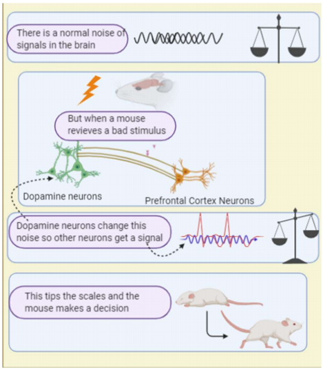
Both phenomena relate to your brain and how dopamine (yes that feel-good chemical in the brain) influences your ability to allow a signal to go through and cause a reaction to a something scary or bad. This is kind of like how you can be in an impressively busy and noisy cocktail party yet, still manage to focus on the person you want to. This filtering is done by screening what is important among the noise. This is, more or less, what happens in the brain too. In a paper recently published in the journal Nature by Weele et. al. on the influence of dopamine and its effect on the medial prefrontal cortex (mPFC: an area in the front of your brain) in changing behaviour. Specifically, behaviour in respect to things we avoid or aversive stimuli. This helps us understand more about the intricate ways dopamine plays with our behaviour and reactions. It also gives us evidence to continue pursuing methods to specifically administer dopamine to targeted areas of the brain instead of all over. The current treatments that involve dopamine distribute it indiscriminately in the brain and can have unintended effects in how we operate. This research helps further this point.
The idea that dopamine modulates behaviour in some way is a long-held notion and has been studied in a variety of situations such as addiction, positive and negative association behavioural mechanisms, visual information discrimination, and working memory.5,6,7,8,9,10,11 It has also been implicated to modulate the mPFC and its activity.1 This is through an area of particularly dense dopaminergic cells called the Ventral Tegmental Area (VTA).2 These dopaminergic cells are cells that secrete dopamine and have further specific characteristics. Before, it had been predicted that dopamine underlays a variety of functions in the mPFC with a role in signal-to-noise ratio.5 A signal-to-noise ratio (SNR) is something like how a radio just plays static if not tuned in properly to a station, or if the signal is not strong enough. However, when a signal is strong enough a sound can be heard on the radio. The same principle applies to the brain. It is constitutively active, especially in processing centres like the mPFC. The result of this is a kind of “static” noise. This would be the noise in the SNR acronym. The signal is when there is some signal from the rest of the body, or other part of the brain, that comes in with information. Now as the mPFC has a lot of areas it effects, it is important to know that it has connections to an area known as the periaqueductal grey (PAG). This is an area of the brain that has been shown to be involved in defensive social behaviours.2,3
In the research conducted by Weele et. al. a variety of methods were used. These involved many surgeries on rats, dissecting and imaging their brains, while they were alive, as well as when they were dead. The scientists used viruses to implant changes to the nerves they were looking at. Well, more than just the nerves in all honesty. It involved the use of fluorescent proteins (proteins that give off light!) and special channel proteins, called rhodopsin channels (these are channels that can be controlled by flashing a specific light). They also used an imaging technique called Fast-scan cyclic voltammetry. This was used to analyse samples after they cut them from the rats’ brains. It is basically, a machine that quickly changes the voltage of a sample, then uses the information gathered to determine the concentration of dopamine. The researchers also performed experiments on the behavior of their rats when conditioned with a sound and pairing it with either a shock or with something delicious. By combining these techniques with some impressive imaging hardware and software the researchers were able to identify some very interesting things.
Among these interesting things was a further link between the VTA and the specific connections between the mPFC and the PAG. The information that this connection codes for is the reaction to an adverse stimulus. What was determined was that this information could just be filtered out as “noise” to a degree if no dopamine was released by the VTA. In order to determine that other areas weren’t changed in their activity; further testing was performed on the mPFC and its connection to an area known as the nucleus accumbens (NA). The NA is an area of the brain that is associated with reward-related processes. The opposite of the PAG in respect to behaviour conditioning. What was found was that even though there were dopamine receptors on the connection between the mPFC and the NA, it appeared the VTA (the specific dopaminergic cells studied) did not change the activity in the NA. What was also surprising was that directly puffing dopamine on the mPFC connection with the PAG didn’t change the SNR. What did change the SNR however, was stimulating the VTA dopaminergic cells. The conclusion to draw here is that the VTA is somehow influencing the signals coming into the mPFC and not only the mPFC itself.
The type of action that would be produced by this connection is also intriguing. Through behavioral tests mice were seen to show a stronger adverse reaction to sounds that signaled the bad stimulus. An electrical shock, in their case, was the bad stimulus. They froze up longer than those mice who did not have their VTA cells stimulated. They also were slower to react when they heard the sound that meant they would get a treat. What can be gleaned is, that dopamine has a specific function that helps us react to things we know are bad or dangerous (see Figure 1), like a thunderclap. When we hear it, we turn to it and freeze, then we act further to preserve our safety.
[+] References
Arnsten, A. F. (2009). Stress signalling pathways that impair prefrontal cortex structure and function. Nature reviews neuroscience, 10(6), 410-422.
Bandler, R., & Carrive, P. (1988). Integrated defence reaction elicited by excitatory amino acid microinjection in the midbrain periaqueductal grey region of the unrestrained cat. Brain research, 439(1-2), 95-106.
Franklin, T. B., Silva, B. A., Perova, Z., Marrone, L., Masferrer, M. E., Zhan, Y., ... & Pagella, S. (2017). Prefrontal cortical control of a brainstem social behavior circuit. Nature neuroscience, 20(2), 260.
Kalivas, P. W. (1993). Neurotransmitter regulation of dopamine neurons in the ventral tegmental area. Brain Research Reviews, 1(1), 75-113.
Kroener, S., Chandler, L. J., Phillips, P. E., & Seamans, J. K. (2009). Dopamine modulates persistent synaptic activity and enhances the signal-to-noise ratio in the prefrontal cortex. PloS one, 4(8).
Noudoost, B., & Moore, T. (2011). Control of visual cortical signals by prefrontal dopamine. Nature, 474(7351), 372-375.
Rebec, G. V., Christensen, J. R., Guerra, C., & Bardo, M. T. (1997). Regional and temporal differences in real-time dopamine efflux in the nucleus accumbens during free-choice novelty. Brain research, 776(1-2), 61-67.
Vander Weele, C. M., Siciliano, C. A., Matthews, G. A., Namburi, P., Izadmehr, E. M., Espinel, I. C., ... & Chang, C. J. (2018). Dopamine enhances signal-to-noise ratio in cortical-brainstem encoding of aversive stimuli. Nature, 563(7731), 397-401.
Venton, B. J., & Wightman, R. M. (2003). Psychoanalytical electrochemistry: dopamine and behavior.
Williams, G. V., & Goldman-Rakic, P. S. (1995). Modulation of memory fields by dopamine Dl receptors in prefrontal cortex. Nature, 376(6541), 572-575.
Zweifel, L. S., Parker, J. G., Lobb, C. J., Rainwater, A., Wall, V. Z., Fadok, J. P., ... & Phillips, P. E. (2009). Disruption of NMDAR-dependent burst firing by dopamine neurons provides selective assessment of phasic dopamine-dependent behavior. Proceedings of the national academy of sciences, 106(18), 7281-7288.
[+] Other Work By Gavin Harvey
I took this new sleeping pill and nhjmjjj...... (it worked)
Neuroanatomy
An Oxford research study, conducted by Kemph, Song, Talbot and Miesenböck, found evidence that the ratio of different states of NADPH had a direct effect on Shaker potassium channels which cause sleeping. This research helps shed light on the mechanisms of sleeping and could help those suffering from insomnia as well as develop better sleeping pills.
Review on the use of 3D cerebral organoids and the current technical progress in their development
Neuroscience In Review