Levels of blood proteins within the brain may be linked to major depressive disorder
A new study found that asymmetry of the frontal areas of the brain was positively correlated with thoughts of suicide in patients with Major Depressive Disorder (MDD).
Author: Ashlynn Dean
Download: [ PDF ]
Neuroanatomy
Although there are many studies over depression and related markers within the brain, there are no studies that specifically test the link between depression and asymmetry in certain brain regions. In a paper recently published by Diagnostics, Seung et al. investigates frontal areas of the brain and how it affects the relationship between depression and suicidal thoughts in patients with MDD.1 This study found that prefrontal asymmetry controlled the effects of depression severity on suicidal thoughts while patients were performing the Verbal Fluency Task (VFT). There was also a positive correlation between prefrontal asymmetry and suicidal thoughts in patients with MDD, along with a lower reported level of asymmetry in patients with MDD compared to those without.
Introduction
Depression is a common mood disorder that can affect the way people think, feel, and perform activities in their daily lives.2 Although this disorder is common, it is also a very serious brain condition that is difficult to diagnose. Some studies have focused on using blood and tissue samples to measure proteins and analyze gene expression.3 Currently, there are no noninvasive, qualitative procedures for diagnosing MDD.4 Fortunately, a recent technique was developed which makes it easier to look at brain activity, called functional near-infrared spectroscopy (fNIRS). This technique is an easy, portable, non-invasive method of monitoring brain activity by detecting a protein within the blood, called Hemoglobin (Hb).5 More specifically, this imaging system tracks changes in the amount of oxygenated (oxy-) and deoxygenated (deoxy-) Hb using light, which then reflects brain activity.1 Although much research is devoted to finding markers within the brain that relate to risk of depression and suicidal thoughts, none have looked at asymmetry within the frontal areas of the brain.
Methods
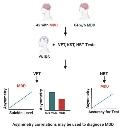
A group of 106 participants were used in this study, 42 with MDD and 64 without. Each participant went through three different cognitive tasks while their Hb levels were measured, including the VFT, Korean Stroop Task (KST), and N-Back Task (NBT). The VFT involves generating as many words with the same first letter as possible in 30 seconds. The KST challenges the patients to choose the correct color of a word when the word itself names a different color. Lastly, the NBT was used to assess working memory by presenting a number (1- 5) every second and requiring the participants to identify the number they saw two numbers prior. Participants filled out a questionnaire to determine personal levels of anxiety, depression, and suicidal thoughts (Scale of 1-4, 1 = absent, 4 = suicide attempts). During these tasks, the participants were to sit still with an fNIRS sensor attached to them. Prefrontal asymmetry was assessed by calculating the changes in Hb between the left and right parts of the brain. The scientists also analyzed the differences between the participants with MDD and their healthy counterparts of similar age and sex (controls).
Results
Throughout the study, no significance was reported for deoxy-Hb, so all results are referring to the asymmetry of oxy-Hb. The study revealed that patients with MDD showed a lower prefrontal asymmetry during the VFT compared to the controls (Figure 1). Patients with MDD also showed a positive correlation between asymmetry during VFT and the selfreported level of suicidal thoughts, while the controls showed a negative correlation between asymmetry and both the KST and anxiety levels (Figure 1). For the NBT, a positive correlation was found between asymmetry and accuracy during the task in MDD participants (Figure 1). Lastly, the VFT indicated a moderation of the relationship between depression severity and suicidal thoughts by prefrontal asymmetry. The moderation of this relationship by prefrontal asymmetry was higher when levels of oxy-Hb were greater in the left side of the brain.
Discussion
These data indicate that asymmetry in the prefrontal regions of the brain may show different levels of depression or suicidal thoughts when performing certain tasks. Currently, the methods for diagnosing MDD and minor forms of depression are not with scientific techniques but rather with checklists and questionnaires.6 Using checklists is not accurate and cannot properly quantify the severity of people’s symptoms. There have been recent updates to the methods of diagnosing and treating depression, including animated systems, but still fall short of long-term treatments for depression.7 Analyzing brain activity using fNIRS is an easy and possibly more effective method of diagnosing depression and suicidal tendencies. The use of fNIRS in neuroscience has grown rapidly in the previous years due to its non-invasive, portable system that is less likely to be affected by bodily movements.8 The use of fNIRS in conjecture with basic cognitive tasks could be a simple, yet promising way of accurately diagnosing depression and risk of suicidal actions. Although this method seems promising, more research is needed on the reliability of the correlation between asymmetry and severity of depression.
[+] References
*Seung, Y. B., Jeong-Youn, K., Jongkwan, C., Ji, Y. B., Yeonsoo, P., Yourim, K., Minjee, J., Seung-Hwan, L. (2019). Prefrontal Asymmetry during Cognitive Tasks and Its Relationship with Suicide Ideation in Major Depressive Disorder: An fNIRS Study. Diagnostics, 9, 193.
National Institute of Mental Health: NIMH (2018, February). Depression. https://www.nimh.nih.gov/health/topics/depression/index.shtml.
Wang, H., Zhang, M., Xie, Q., Yu, J., Qi, Y., Yue, Q. (2019). Identification of diagnostic markers for major depressive disorder by cross-validation of data from whole blood samples. PeerJ, 7, e7171.
Hacimusalar, Y. & Esel, E. (2018). Suggested Biomarkers for Major Depressive Disorder. Noropsikiyatri Arsivi, 55(3), 280-290.
Irani, F., Platek, S. M., Bunce, S., Ruocco, A. C., Chute, D. (2007). Functional near infrared spectroscopy (fNIRS): an emerging neuroimaging technology with important applications for the study of brain disorders. Clinical Neuropsychology, 21(1), 9-37. doi: 10.1080/13854040600910018.
Thomas-MacLean, R., Stoppard, J., Miedema, B., Tatemichi, S. (2005). Diagnosing depression. Canada Family Physicians, 51(8), 1103.
Sanchez, K., Eghaneyan, B. H., Trivedi, M. H. (2016). Depression Screening and Education: Options to Reduce Barriers to Treatment (DESEO): protocol for an educational intervention study. BMC Health Services Research, 16, 322.
Pinti, P., Tachtsidis, I., Hamilton, A., Hirsch, J., Aichelburg, C., Gilbert, S., Burgess, P. W. (2020). The present and future use of functional near-infrared spectroscopy (fNIRS) for cognitive neuroscience. Annuals of the New York Academy of Sciences, 1464(1), 5-29.
[+] Other Work By Ashlynn Dean
A study of neuronal distribution within the brain of epileptic patients shows the preservation of a certain neuron
Neurophysiology
A new study finds a link between distribution patterns of neurons within the brain and tissue samples from epileptic patients, indicating that a certain interneuron is mainly preserved in the epileptic brain.
Review of Drug Addiction and the Role of Perineuronal Nets
Neuroscience In Review