Use it or Lose It: How Bilingualism May Help Prevent Cognitive Decline
Author: Isabella Moreno Stedman
Neuroanatomy
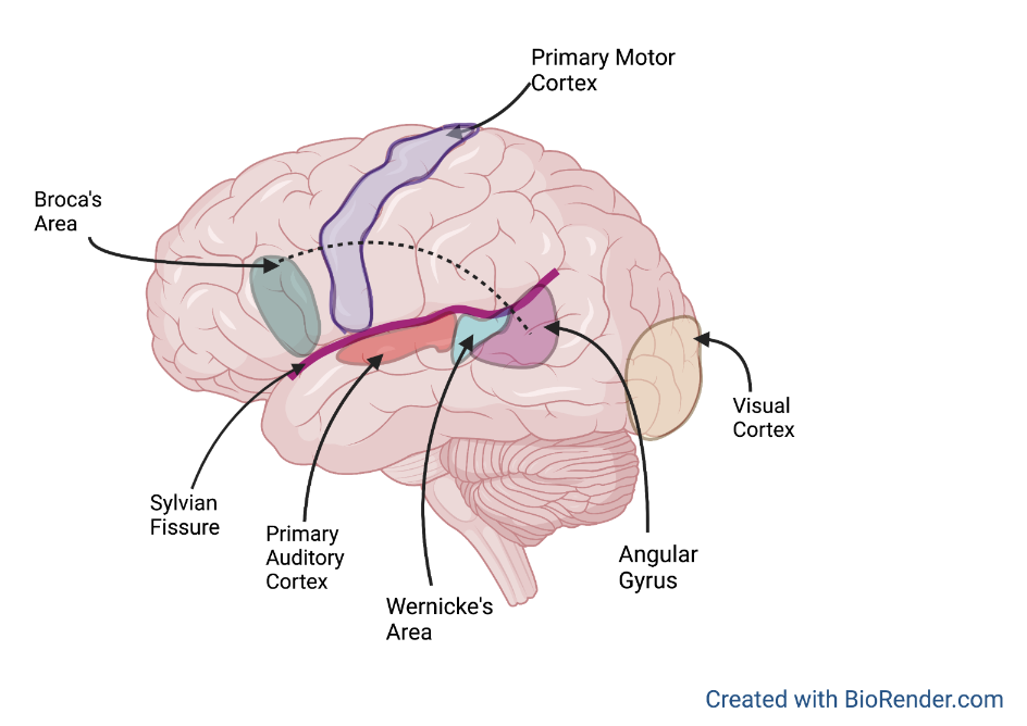
The United States has long been considered a monolingual country, now most people over the age of five have reported speaking more than one language or a language other than English at home (Marian, 2012). This increase in bilingualism not only benefits cross-cultural communication but may also have cognitive benefits. Despite this large increase in bilingual individuals, little is known about how bilingualism affects the structure of the brain. It is known that the components of language are processed by a designated area of the cortex called the perisylvian language zone or “house of speech” (Catani, 2005).
Despite this knowledge few studies have been done to investigate if proficiency and age of acquisition of the second language affects brain structure and if these changes affect age related cognitive decline. An article published in 2021 in the Journal of Anatomy investigated the role of speaking multiple languages has on brain structure. This study found that neuroanatomical changes are found in individuals who speak multiple languages, and the degree of this change is influenced by proficiency and age of acquisition of the second language. Evidence also showed that bilingualism reduces the likelihood and symptoms of age related cognitive decline. This study is unique in that it provides a comprehensive examination of factors that may cause these structural changes and highlights results from numerous studies in this area of research. In this way the study aims to help further the understanding of how language affects brain structure and overall cognitive health which may help prevent diseases and decrease severity of symptoms.
The brain areas responsible for language processing and composition have long been accepted. The function of a brain area can often be discovered by observing what occurs when these structures are damaged or missing. Damage to specific structures within this area causes a variety of symptoms including spelling deficits (Henry, 2007). Damage to these areas may also result in a condition known as aphasia which is the loss of the ability to understand or express speech, and a decline in language components such as grammar or syntax (Le, 2021). Various imaging techniques can be used to visualize brain structures and activity during tasks, these help improve the understanding of the composition of these language related areas of the brain. Previous hypotheses known as the “Critical Period Hypothesis”, have suggested that the proficiency acquired when learning a native language cannot be rivaled when learning a second later in life. When people are younger and learning their native language, the brain more readily forms strong connections that allow for proficient language processing and comprehension. Later in life, the brain requires more practice or repetition to strengthen these connections, therefore second languages do not develop as easily or as well after 12 years old (Vanhove, 2013). Other hypotheses proposed that the studies argue that language acquisition and comprehension depend on memory and consciousness during the learning process. Both hypotheses have since been challenged and it is now argued that second language acquisition does not have a “critical” period or age when it most successfully occurs.
This study was done by conducting various keyword searches for published articles using scientific databases. Searches were focused on articles that discussed structural changes due to language and diseases. The study was based on multiple imaging studies and used imaging techniques to observe anatomical similarities and differences. Researchers were able to observe patterns of activity to determine what areas of the brain were active during specific speech tasks. These articles were screened so that only relevant information was included.
Results indicate that individuals who are highly proficient in their native and second language display a wide overlap of brain structures involved in language processing. Other areas were found to overlap regardless of proficiency. Imaging results revealed that second language acquisition occurs through the same brain connections as the first language (Optiz, 2004). Imaging studies show that there is more brain activity during conjugation and sentence composition while speaking the second language. Not only is more brain activity required to maintain a second language but also to inhibit one language while the other is in use.
The cortex is the outermost portion of the brain and is composed of gray and white matter (Jawabri, 2021). Gray matter is composed of brain cell bodies that function to receive incoming information and regulate outgoing information. White matter is responsible for transmitting this information back and forth (Wen, 2005). Increase in gray and white matter density in various language related brain regions has also been found in people who are bilingual. This was found by comparing cortical thickness of monolinguals and bilinguals. How these anatomical differences affect disease is not fully understood. This study also mentions there is a correlation between bilingualism and an increase in brain cell regeneration. This may be because the production of new brain cells occurs in the same structure that is responsible for multiple memory components, some of which are required to recall words or concepts (Van de Ven, 2020).
This study is important and provides valuable information as it consolidates many theories, hypotheses, and evidence on how the brain changes in response to bilingualism. This is important due to the recent increase in bilinguals. These structural changes due to bilingualism result in an increase in executive function, and capacity for conflict resolution. The degree of structural changes is dependent on age of acquisition and proficiency with younger ages of acquisition and higher proficiency being correlated with increased structural changes. Current evidence suggests that proficiency has a greater impact than age of acquisition does. This study is also important as it provides evidence on how to prevent and offset symptoms of age-related cognitive decline. Understanding age-related cognitive decline is important to our elderly population since age related diseases increase the rate at which brain cells die and tissue is lost (Murman, 2015). This loss can lead to the inability to complete daily tasks and result in loss of independence.
[+] References
Marian, V., & Shook, A. (2012). The cognitive benefits of being bilingual. Cerebrum : the Dana forum on brain science, 2012, 13.
- Catani, M., Jones, D. K., & ffytche, D. H. (2005). Perisylvian language networks of the human brain. Annals of neurology, 57(1), 8–16. https://doi.org/10.1002/ana.20319
Henry, M. L., Beeson, P. M., Stark, A. J., & Rapcsak, S. Z. (2007). The role of left perisylvian cortical regions in spelling. Brain and language, 100(1), 44–52. https://doi.org/10.1016/j.bandl.2006.06.011
Le, H., & Lui, M. Y. (2021). Aphasia. In StatPearls. StatPearls Publishing
Vanhove J. (2013). The critical period hypothesis in second language acquisition: a statistical critique and a reanalysis. PloS one, 8(7), e69172. https://doi.org/10.1371/journal.pone.0069172
Bertram Opitz and Angela D. Friederici. Brain Correlates of Language Learning: The Neuronal Dissociated of Rule-Based vs. Similarity Based Learning. Journal of Neuroscience 29 September 2004, 24 (39) 8436-8440; DOI: https://doi.org/10.1523/JNEUROSCI.2220-04.2004
Jawabri, K. H., & Sharma, S. (2021). Physiology, Cerebral Cortex Functions. In StatPearls. StatPearls Publishing.
Wen, Q., & Chklovskii, D. B. (2005). Segregation of the brain into gray and white matter: a design minimizing conduction delays. PLoS computational biology, 1(7), e78. https://doi.org/10.1371/journal.pcbi.0010078
Van de Ven, V., Waldorp, L., & Christoffels, I. (2020). Hippocampus plays a role in speech feedback processing. NeuroImage, 223, 117319. https://doi.org/10.1016/j.neuroimage.2020.117319
Murman D. L. (2015). The Impact of Age on Cognition. Seminars in hearing, 36(3), 111–121. https://doi.org/10.1055/s-0035-1555115