Sleep Less, Remember Less
Researchers have shown that the dendritic spine density in the hippocampus, a structure important for memory, was increased with lack of sleep which is related to weaker memory consolidation.
Author: Andrea Nguyen
Download: [ PDF ]
Neuroanatomy
A paper published in 2019 by Barbara Gisabella and associates examined how sleep, more specifically how the lack of sleep, effected the branches of neurons in the hippocampus. This research allowed for insight needed to understand how important sleep is and its overall effects on the dendritic spines of neurons in the hippocampus because these are important for memory. This study primarily found that sleep deprivation causes an increase in these neuron branches while sleep does the opposite (Gisabella et al., 2019). These findings are important as they imply that the brain consolidates memories better during sleep which causes these dendritic spines to decrease. Therefore, it is implied that sleep deprivation does the opposite as the brain cannot perform this process in times of long wakefulness. These findings are also important for future studies in emotional memory consolidation because emotional memories are seen to be stronger than regular memories. Therefore, it can be said that the dendritic spines would change with sleep deprivation for emotional memories based off the results of this study.
Background
Sleep is a process essential for healthy living as it effects brain functionality and physiology within the body (Medic et al., 2017). The specific functions of sleep have been widely researched for decades, but one important function that has been determined is its essentiality for memory consolidation (Worley, 2018). In a study done by Laurel Graves (2003) and colleagues it was observed that sleep deprivation caused damage to memory consolidation within the hippocampus. Therefore, an important structure for this process during sleep is the hippocampus (Havekes et al., 2016). The hippocampus is a s-shaped structure that is referred to as a limbic lobe due to its location in the temporal area of the cerebral cortex (Anand & Dhikav, 2012). In terms of memory consolidation, it was observed that the CA1 region of the hippocampus plays an important role, due to inactivation of this area causing disruption in contextual memory (Daumas et al., 2005).
Therefore, memory consolidation in this area is thought to rely on the strength of specific synapses, which means that the size and amount dendritic spines play an important role (Gisabella et al., 2019). Moreover, it was expected that contextual memory consolidation would cause changes in CA1 dendritic spines (Gisabella et al., 2019). A previous study done by Lusia de Vivo (2017) and colleagues found that the synapses of the spine density was decreased during sleep and increased in periods of sleep deprivation (de Vivo et al., 2017). Therefore, this is an important finding for understanding the overall effects of sleep deprivation on dendritic spines. However, there have been results that contradict these findings. Therefore, this study sought to understand this process further by observing the overall alterations of the dendritic spines in the CA1 for sleep deprivation and normal sleep.
Methods
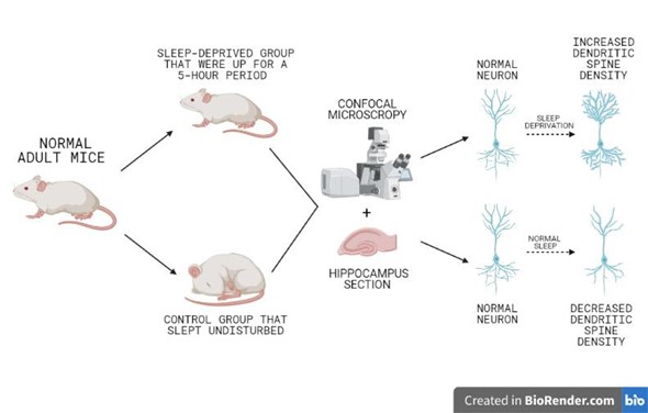
This study was conducted on adult mice that were injected with a chemical used to target the CA1 of the dorsal hippocampus (Giasbella et al., 2019). They were randomly split into a sleep deprivation group and a control group (Gisabella et al., 2019). Sleep deprivation was done by keeping a light on in a controlled cage room for 5 hours (7 AM-12PM); the experimenters also gently shook the cage if they saw any mice dozing off (Gisabella et al., 2019). The control mice were left alone in their individual cages and were able to sleep normally (Gisabella et al., 2019). After the 5-hour period the mice were anesthetized and their brains were surgically removed and cut into sections (Gisabella et al., 2019). These sections were used to determine the density of the dendritic spines using confocal microscopy (Gisabella et al., 2019). Confocal microscopy is when the light of the microscope is aimed on a small specific area inside the tissue, instead of the entire area of the sample (Nwaneshiudu et al., 2012). This process was paired with an imaging software allowing 3D images of the dendritic spines in the CA1 neurons for both the controlled and sleep deprived mice which were then quantified (Gisabella et al., 2019). The examined dendritic spine density was located on the proximal and distal areas of the sections and they primarily observed the apical and basal dendrites (Gisabella et al., 2019).
Results
Dendritic spine density was significantly increased in the mice that were sleep deprived in comparison to the control mice (fig.1) (Gisabella et al., 2019). It was also observed that the volume of the spines was also greater in the sleep deprived mice (Gisabella et al., 2019). The primary branches of the dendritic spines were greater in the sleep deprived mice, though not in the secondary branches (Gisabella et al., 2019). It was also observed that the density of the apical dendrites in the distal area was greater in the sleep deprived mice (Gisbella et al., 2019). The density of both apical and basal dendrites was greater in the proximal area in the sleep deprivation mice (Gisbella et al., 2019). Moreover, the apical dendrites in the stratum radiatum of the sleep deprived mice was increased as was the basal dendrites in the stratum oriens (Gisabella et al., 2019).
Conclusion
Based off the results of this study it can be said that sleep deprivation causes an increase in dendritic spines and sleep causes a decrease (fig.1) (Gisabella et al., 2019). This means that during sleep the brain can renormalize synaptic strength and consolidate memories which means the density of the dendritic spine synapses are decreased (Spano et al., 2019). Therefore, it can be said that sleep deprivation has an opposite effect and prevents memory consolidation within the hippocampus as the brain cannot renormalize these synapses. This provides explanation to why the hippocampus is not able to effectively consolidate memory after sleep deprivation. Moreover, by understanding this relationship future studies can evaluate how this relationship effects consolidation of emotional memories. It has been observed that emotional memories, especially ones correlated with negative stimuli, have a greater rate of recollection and recognition (Harrington et al., 2018). One example of this emotional memory is fear memory which is widely studied. Therefore, further research could study the changes of dendritic spine behavior after periods of sleep deprivation and sleep following an emotionally charged behavior.
[+] References
Anand, K. S., & Dhikav, V. (2012). Hippocampus in health and disease: An overview. Annals of Indian Academy of Neurology, 15(4), 239–246. https://doi.org/10.4103/0972-2327.104323.
Daumas, S., Halley, H., Francés, B., & Lassalle, J. M. (2005). Encoding, consolidation, and retrieval of contextual memory: differential involvement of dorsal CA3 and CA1 hippocampal subregions. Learning & memory (Cold Spring Harbor, N.Y.), 12(4), 375–382. https://doi.org/10.1101/lm.81905.
de Vivo, L., Bellesi, M., Marshall, W., Bushong, E. A., Ellisman, M. H., Tononi, G., & Cirelli, C. (2017). Ultrastructural evidence for synaptic scaling across the wake/sleep cycle. Science (New York, N.Y.), 355(6324), 507–510. https://doi.org/10.1126/science.aah5982.
Genzel, L., Rossato, J. I., Jacobse, J., Grieves, R. M., Spooner, P. A., Battaglia, F. P., Fernández, G., & Morris, R. G. (2017). The Yin and Yang of Memory Consolidation: Hippocampal and Neocortical. PLoS biology, 15(1), e2000531. https://doi.org/10.1371/journal.pbio.2000531.
Gisabella, B., Scammell, T., Bandaru, S. S., & Saper, C. B. (2019). Regulation of hippocampal dendritic spines following sleep deprivation. Journal of Comparative Neurology, 528(3), 380–388. https://doi.org/10.1002/cne.24764.
Graves, L. A., Heller, E. A., Pack, A. I., & Abel, T. (2003). Sleep deprivation selectively impairs memory consolidation for contextual fear conditioning. Learning & memory (Cold Spring Harbor, N.Y.), 10(3), 168–176. https://doi.org/10.1101/lm.48803.
Harrington, M. O., Nedberge, K. M., & Durrant, S. J. (2018). The effect of sleep deprivation on emotional memory consolidation in participants reporting depressive symptoms. Neurobiology of Learning and Memory, 152, 10–19. https://doi.org/10.1016/j.nlm.2018.04.013.
Medic, G., Wille, M., & Hemels, M. E. (2017). Short- and long-term health consequences of sleep disruption. Nature and science of sleep, 9, 151–161. https://doi.org/10.2147/NSS.S134864.
Nwaneshiudu, A., Kuschal, C., Sakamoto, F.H., Anderson, R.R., Schwarzenberger, K., & Young, R.C. (2012). Introduction to Confocal Microscopy. Journal of Investigative Dermatology, 132(12), P1-5. https://doi.org/10.1038/jid.2012.429.
Spano, G. M., Banningh, S. W., Marshall, W., de Vivo, L., Bellesi, M., Loschky, S. S., Tononi, G., & Cirelli, C. (2019). Sleep Deprivation by Exposure to Novel Objects Increases Synapse Density and Axon–Spine Interface in the Hippocampal CA1 Region of Adolescent Mice. The Journal of Neuroscience, 39(34), 6613–6625. https://doi.org/10.1523/jneurosci.0380-19.2019.
Worley S. L. (2018). The Extraordinary Importance of Sleep: The Detrimental Effects of Inadequate Sleep on Health and Public Safety Drive an Explosion of Sleep Research. P & T: a peer-reviewed journal for formulary management, 43(12), 758–763.