Stimulation frequency contributes to reward and aversion
New findings in the continued effort to understand the neuropsychiatry of addiction and depression suggest stimulation patterns in neuronal reward pathways contribute to reward and aversion behaviors.
Author: Trent Pratt
Download: [ PDF ]
Neurophysiology
There are many challenges one must face in the world, and we must adapt our behaviors to receive rewards in order to benefit from opportunities and to avoid punishments and cope with potential disappointments. There are many common clinical conditions such as depression and addiction that have raised questions regarding potential treatments and solutions to improve the quality of life in those that may struggle with these issues. Several regions in the brain are involved in someone’s ability to process reward and aversive behaviors in response to a given situation. Deficits in the brain’s ability to process reward and aversion can lead to the onset of several psychiatric disorders, i.e. depression and addiction. The prevalence of these conditions has raised questions about what physiological dysfunction may be present in the nervous system, and more importantly, how to solve it. It is important to attempt to answer some of these questions and understand how the patterns of specific nervous system cells differ in rewarding and aversive tasks in order to better assess how the two subpopulations of cells work together to ultimately generate behavior.
A paper recently published in Molecular Psychiatry by Carina Soares-Cunha and colleagues investigated the roles of medium spiny neuron (MSN) subtypes in the nucleus accumbens (NAc) of the ventral striatum and their respective roles in reward and aversion.1 MSNs can be subtyped by the types of dopamine receptors they contain: D1-MSNs and D2-MSNs.2 MSNs bind dopamine using D1 receptors (makes them more active) while some bind dopamine by expressing D2 receptors (decreases their activity). A complication in understanding the connectivity of the striatum arises because MSNs are inhibitory neurons and reducing their activity increases the activity of the cells they inhibit. There are numerous studies attempting to understand the relationship between these two subpopulations and suggest they play a major role in reward and aversive behaviors.3,4,5 This study sought to further investigate the relationship between these two striatal neuron subtypes by measuring differences in stimulation frequencies and the resulting effects on reward and aversion.
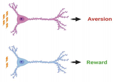
The primary findings of the study were that D1- and D2-MSNs can influence reward and aversion. Stimulation patterns of NAc D1- or D2-MSNs at the cell nucleus, the soma, and at the terminal, the synapse, induced reward or aversion, depending on the characteristics of MSN frequency of stimulation. Positive reinforcement, i.e. reward, was induced by briefly stimulating the neurons. In contrast, aversion was induced by prolonged stimulation of both MSN subtypes. The difference in stimulation patterns resulted in divergent downstream pathways in the VTA and the VP, referring to their impact on either reward or aversive behaviors.
It is important for the brain to determine which environmental situations and stimuli are rewarding and those that are not. The manner in which the brain processes reward and motivational situations is by encoding reward and aversion, where positive reinforcement occurs with reward, and negative reinforcement with aversion. The brain processes information through distinct pathways, in this case, the mesolimbic reward pathway, which is crucial in understanding psychiatric disorders such as depression and addiction.6 The striatum plays a crucial role in this circuitry, particularly the NAc.7 The NAc is a key player in the brain’s ability to process and respond to rewarding and aversive behaviors. There are a number of factors that can affect this region’s typical function, such as chronic stress and gene expression.2 When dysfunction does occur, an individual may be vulnerable to depressive and addictive behaviors, so it is important to understand the relationship between neurons in this region. MSNs in this region different receptors for dopamine, expressing either D1 dopamine receptors, which induce facilitation or D2 dopamine receptors, which induce inhibition.8 The relationship between the activity of these two striatal subtypes determines the effects on reward-related behavior. The implications of this research are important in the continued effort to understanding how the brain processes rewards and their effects on behavior. The resulting behaviors are crucial for the maintenance of an individual’s success that could determine the quality of life of a given individual, or even the survival of an individual or an organism.
There is a bit of controversy attempting to explain the roles of D1- and D2- MSNs and the effects on striatal function and ultimately reward-related behaviors. This study is an attempt to answer some of the questions and proposes that the opposition that has existed in the field should be further explored. In the striatum, the majority of neurons, upward of 95%, are MSNs.9 Both subtypes of MSNs are GABAergic, meaning they are inhibitory and release the neurotransmitter GABA. There are numerous neurotransmitters, i.e. dopamine, that bind to a single receptor, in this case, either D1- or D2- MSNs, and vary in their effects on reward and aversion.10 MSNs receive excitatory input via glutamate on the heads of the numerous spine synapses on the ends of neurons, dendrites, and dopamine inputs are received at the necks of these dendritic spiny synapses.11 The combined activity from both glutamatergic and important neurotransmitters ultimately determines the activity of a given MSN, which by extension, would determine an individual’s behavioral response.
In this study, mice were optogenetically modified to express a light-activated channel which allowed the researchers to stimulate specific sets of inputs onto MSNs using blue light. This optogenetic approach injected mice with halorhodopsin (activated under blue light), which fluxes Cl- which allows light to hyperpolarize and inhibit neurons; channelrhodopsin (activated under amber light), which fluxes Na+ which depolarizes cells to excite them; and eYFP for the control group. Historically, there has been some controversy regarding which NAc MSN subpopulation encodes reward and aversion, so this study used optogenetic manipulation to target NAc D1- or D2-MSN activity using this approach to allow for greater specificity of MSN activity. It involved placing an electrode to stimulate the neurons with a fiber optic patch in the region being studied, in this case, the NAc. Stimulation of individual neurons were then recorded and statistically analyzed. This study found that brief stimulation resulted in increased ventral tegmental area VTA dopaminergic activity either directly, for D1-MSNs alone, or indirectly via the VP for both D1- and D2-MSNs. In contrast, the study also found that prolonged stimulation of the two MSN subpopulations resulted in varying effects on activity downstream, indicating that D1- and D2- MSNs’ stimulation patterns are implicated in both reward and aversive behaviors. Additionally, optogenetic inhibition of D1-MSNs induced aversion and similar results were found in D2-MSN optical inhibition.
This study found that brief stimulation resulted in increased ventral tegmental area VTA dopaminergic activity either directly, for D1-MSNs alone, or indirectly via the VP for both D1- and D2-MSNs. In contrast, the study also found that prolonged stimulation of the two MSN subpopulations resulted in varying effects on activity downstream, indicating that D1- and D2- MSNs’ stimulation patterns are implicated in both reward and aversive behaviors. Additionally, optogenetic inhibition of D1-MSNs induced aversion and similar results were found in D2-MSN optical inhibition.
The hypothesis of this study was to determine the relationship D1- and D2-MSN activity has on reward and aversive behaviors by changing activation patterns of these two striatal neuron types, as well as changes in regions receiving input from projecting MSNs, the VTA (D1-MSNs only) and VP (both). Because this study finds that the two subtypes of MSNs have distinct and varying effects on rewardrelated behaviors, it suggests there is a complex relationship that warrants more research in the field. In doing so, the search for answers in treating psychiatric disorders such as depression and addiction could potentially find a better direction. This study sheds light on what appears to be a controversial area of research, suggesting that both subtypes can drive reward and aversion, determined by their pattern of stimulation. The methodology via optogenetic manipulation allowed for specific neurons to be targeted, versus stimulation the local region of other methods, which does not allow for as much specificity. In doing so, it provided some important findings in the relationship between the activities of striatal MSNs. The major takeaway from this research is that striatum is organized in a more complex manner than was previously thought, and additional research can help shed more light on the matter, which could lead to more potential solutions in addressing psychiatric disorders due to striatal dysfunction.
[+] References
Soares-Cunha, C., Vasconcelos, N. A. P. D., Coimbra, B., Domingues, A. V., Silva, J. M., Loureiro-Campos, E., … Rodrigues, A. J. (2019). Nucleus accumbens medium spiny neurons subtypes signal both reward and aversion. Molecular Psychiatry. doi: 10.1038/s41380-019-0484-3
Russo, S. J., & Nestler, E. J. (2013). The brain reward circuitry in mood disorders. Nature Reviews Neuroscience, 14(9), 609–625. doi: 10.1038/nrn3381
Cui, G., Jun, S. B., Jin, X., Pham, M. D., Vogel, S. S., Lovinger, D. M., & Costa, R. M. (2013). Concurrent activation of striatal direct and indirect pathways during action initiation. Nature, 494(7436), 238–242. doi: 10.1038/nature11846
Natsubori, A., Tsutsui-Kimura, I., Nishida, H., Bouchekioua, Y., Sekiya, H., Uchigashima, M., … Tanaka, K. F. (2017). Ventrolateral Striatal Medium Spiny Neurons Positively Regulate Food-Incentive, Goal-Directed Behavior Independently of D1 and D2 Selectivity. The Journal of Neuroscience, 37(10), 2723–2733. doi: 10.1523/jneurosci.3377-16.2017
Vicente, A. M., Galvão-Ferreira, P., Tecuapetla, F., & Costa, R. M. (2016). Direct and indirect dorsolateral striatum pathways reinforce different action strategies. Current Biology, 26(7). doi: 10.1016/j.cub.2016.02.036
Nestler, E. J., & Carlezon, W. A. (2006). The Mesolimbic Dopamine Reward Circuit in Depression. Biological Psychiatry, 59(12), 1151–1159. doi: 10.1016/j.biopsych.2005.09.018
Schultz, W. (2000). Multiple reward signals in the brain. Nature Reviews Neuroscience, 1(3), 199–207. doi: 10.1038/35044563
Chuhma, N., Tanaka, K. F., Hen, R., & Rayport, S. (2011). Functional Connectome of the Striatal Medium Spiny Neuron. Journal of Neuroscience, 31(4), 1183–1192. doi: 10.1523/jneurosci.3833-10.2011
Kemp, J.M & Powell T.P. (1971). Philosophical Transactions of the Royal Society of London. B, Biological Sciences, 262(845), 441–457. doi: 10.1098/rstb.1971.0106
Tejeda, H. A., Wu, J., Kornspun, A. R., Pignatelli, M., Kashtelyan, V., Krashes, M. J., … Bonci, A. (2017). Pathway- and Cell-Specific Kappa-Opioid Receptor Modulation of Excitation-Inhibition Balance Differentially Gates D1 and D2 Accumbens Neuron Activity. Neuron, 93(1), 147–163. doi: 10.1016/j.neuron.2016.12.005
Xu, Z., Wilson, C., & Emson, P. (1989). Restoration of the corticostriatal projection in rat neostriatal grafts: electron microscopic analysis. Neuroscience, 29(3), 539–550. doi: 10.1016/0306-4522(89)90129-2
[+] Other Work By Trent Pratt
Musical enrichment for neurologically disordered children
Neuroanatomy
A new study finds music therapy an effective addition to treatment for children with severe neurological disorders, enhancing the children’s attentional and emotional capacities.
An overview of exercise-induced hippocampal neurogenesis
Neuroscience In Review