Size does matter
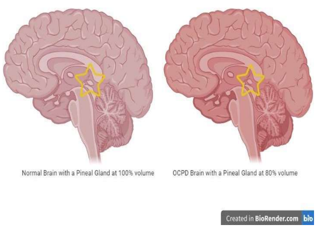
The article discusses the difference in the size of the pineal gland between those with obsessive-compulsive personality disorder (OCPD) and those without it. The size difference matters as those with smaller pineal gland have consistently been diagnosed with OCPD.
Author: Cori Dougher
Download: [ PDF ]
Neuroanatomy
The motivation behind this research is that our knowledge of the pathology of personality disorders is not very vast, especially regarding OCPD. Our knowledge of personality disorders is limited to psychoanalytical approaches alone. This is why performing the brain imaging of those with OCPD is crucial to gain knowledge on the biological aspects of this personality disorder to better understand how this disorder comes about. In the study by Murad Atmaca and Colleagues, they studied the size of the pineal gland of patients diagnosed with OCPD and healthy subjects by sending them through magnetic resonance imaging (MRI) and comparing the images of these patients. The findings showed that those with OCPD have a pineal gland volume 20% smaller than the normal patients. Also, to note was that hormonal changes of the pineal gland by melatonin and pineal calcifications were not included in the volumes of the pineal gland size, which could skew the results in a different way if these were considered. The findings of the study still show that those diagnosed with OCPD have significantly smaller pineal glands. This could prove that OCPD falls on the OCD spectrum of disorders due to similar results of MRI scans.
This study was based on other research done by Atmaca and colleagues. In an unpublished study done by Atmaca and colleagues, their team examined the size of the orbitofrontal cortex or the OFC, along with the thalamus in patients with and without OCPD. The study found that those who had been diagnosed with OCPD their MRI image showed much smaller left and right OFC’s compared to those of normal patients, but those with OCPD had a greater size thalamus than those of normal patients. This unpublished study by Atmaca is what drove them to also study the pineal gland of those same patients that are normal and those that are diagnosed with OCPD.
The pineal gland, as stated by Acer in “Pineal Gland and Melatonin: Recent Advances in Development, Imaging, Disease and Treatment” is a small, pinecone shaped organ of the brain that has a part in the circadian rhythms and sleep cycles by secreting melatonin at the appropriate times. The secretion of melatonin is altered in those with schizophrenia, bipolar disorder, and major depressive disorder as mentioned by Brown and their colleagues in their study on melatonin secretion. Catapano and Monteleone performed a study to track the melatonin release in normal patients and Obsessive-compulsive disorder or OCD over a 24- hour period. The patients with OCD had abnormal secretion of melatonin leading to troubles falling and staying asleep. This study was the other driver of the study by Atmaca as the pineal gland size was observed in OCD patients and normal patients and it was noted that those with OCD had a pineal gland volume that of 80% of the volume in comparison with the normal patients. This led Atmaca to think about how pineal gland sizes differ in those with not only OCD but OCPD as well.
The group utilized for this study was composed of consenting patients who were diagnosed with obsessive-compulsive personality by the clinics of the Firat University School of medicine Department of Psychiatry as well as ones diagnosed as healthy. These patients were between the ages of 18-65 and also participated in the previous study by Atmaca where they studied the size of the thalamus and the OFC in OCPD and normal patients. There was a normal patient for every diagnosed patient by matching based on age, sex, education, and handedness. If any of these patients were found to have any other psychiatric disorders, medical problems, malformations of the brain, or any substance abuse within 6 months of the study they were emitted from the study to prevent any discriminations. The procedure consisted of taking MRI images of the pineal gland for both the diagnosed and normal patients. The pictures were taken by a neuroradiologist researcher who was ignorant of who had OCPD and who was normal along with being verified by a second researcher who was ignorant as well. The boundaries of the pineal gland were determined by Jackson, Patel, Yuh, Sumida, Findikli, and their colleagues which were the superior colliculus, the quadrigeminal cistern, and the posterior portion of the third ventricle. Sagittal, Coronal, and Axial views were all taken of the pineal gland to see its entirety. The height and width of the pineal gland were measured on the coronal images and the length was measured on the axial images. These images were then transferred to a semi-automated program to obtain the volumetric results. To perform statistical analysis, the statistical package for social sciences was used. A t-test was used to compare the mean ages and volumetric differences between the patients with OCPD and the normal patients. A chi-square test was done to assess the gender distribution among the patients. A volumetric comparison of age, gender, and total brain volume was performed. The last statistical analysis to be performed was the Spearman’s correlation test which detects correlations between pineal gland volumes and the demographic/clinical parameters as well.
Analyzing the demographics of the 16 patients with OCD and the 18 patients in the control group (the normal patients) showed they are pretty similar overall, which was the intent so it wouldn’t be a major factor. The age of the OCPD group averaged 32.5 years old plus or minus 8.9 years while the average age for the control group is 29.5 years old plus or minus 5.1 years. There is a ratio of 11 females to 5 males in the OCPD group while there are 10 females and 8 males. For the OCPD patients, 9 of them at least finished high school while 15 from the normal group finished high school, while the rest from both groups (7 and 3 respectively) did not. Everyone from both groups were right-handed. On the depression rating scale, the OCPD group had an average score of 9.8 plus or minus 2.2 points while the normal group had an average score of 3.3 plus or minus 1.7 points. The OCPD patients had been diagnosed with their illness on average about 6.4 years from the start of the study plus or minus 2.2 years.
While there were no differences in the gray and white matter volumes of the brain between the two groups, the volumes of the pineal glands differed greatly. The average size of the pineal gland in the group diagnosed with OCPD was 20% smaller than the normal group. Even when controlling for gender, age, and overall brain volumes, those diagnosed with OCPD still had significantly smaller pineal gland volumes. When utilizing a correlation test, there was no correlational relationship between any of the demographical or clinical variables and pineal gland volumes among the OCPD patients and normal patients.
This was the first study on the volumes of the pineal gland in those diagnosed with OCPD so the information gathered from this study can help to grasp a better understanding of OCPD. While OCPD is normally classified as a personality disorder there has been increasing evidence stated by Fineberg and colleagues that it could actually be more like a neurocognitive function disorder. Stein states how OCPD when in comparison with OCD based on phenomenology, comorbidity, heritability, risk factors, the course of the illness, and response to treatment shows OCPD falls among personality disorders, obsessive-compulsive, and related disorders, which is conflicting with the previous statement by Fineberg. This study done by Atmaca and a previous study of theirs also listed in this paper, shows that those with OCPD has significantly reduced sizes of OFC, the thalamus, and the pineal gland, which was also found to be true in patients with OCD in previous studies done by Atmaca. The sum of these studies shows that OCPD and OCD could fall onto the same spectrum of disorders based on neuroanatomy. While normally studying such a small sample size would not permit these drastic of claims, since OCPD normally has comorbid situations it can be hard to perform studies on those with OCPD so this small sample size is acceptable. Considering all of this, I believe further studies are necessary to draw conclusions that could change the way we associate and diagnose OCPD. While OCD and OCPD are similar in nature, I believe this study does prove that they fall within the same spectrum of mental disorders, but a study with a larger sample size is necessary to draw any further conclusions.
[+] References
Acer N, Turgut M, Yalcin SS, Duvernoy HM. Anatomy of the human pineal gland. In: Turgut M, Kumar R, editors. Pineal Gland and Melatonin: Recent Advances in Development, Imaging, Disease and Treatment. Nova Science; 2011.
Atmaca M, Korucu T, Tabara MF, Yildirim H, Kılıc MC. Volumetric MRI study of orbito- frontal cortex and thalamus in obsessive-compulsive personality disorder. J Clin Neurosci 2019;64:89–93.
Atmaca M, Yildirim H, Ozdemir H, Aydin A, Tezcan E, Ozler S. Volumetric MRI assessment of brain regions in patients with refractory obsessive-compulsive disorder. Prog Neuropsychopharm Biol Psych 2006;30(6):1051–7.
Atmaca M, Yildirim H, Ozdemir H, Tezcan E, Poyraz AK. Volumetric MRI study of key brain regions implicated in obsessive–compulsive disorder. Prog Neuropsychopharm Biol Psych 2007; 31(1):46–52.
Brown R, Kocsis JH, Caroff S, Amsterdam J, Winokur A, Stokes PE, et al. Differences in nocturnal melatonin secretion between melancholic depressed patients and control subjects. Am J Psych 1985; 142:811–6.
Catapano F, Monteleone P, Fuschino A, Maj M, Kemali D. Melatonin and cortisol secretion in patients with primary obsessive-compulsive disorder. Psych Res 1992; 44(3):217–25.
Fineberg NA, de Koenigswarter N, Reghunandanan S, Kolli S, Jefferies K, Laws K. The neurocognitive profile of obsessive compulsive personality disorder; a preliminary analysis. Barcelona Spain: Abstract for a poster ICOCS annual scientific meeting, 2013.
Findikli E, Inci MF, Gokce M, Findikli HA, Altun H, Karaaslan MF. Pineal gland volume in schizophrenia and mood disorders. Psych Danub 2015; 27(2):153–8.
Jackson GD, Duncan JS. MRI neuroanatomy: a new angle on the brain. WB Saunders Company; 1996.
Monteleone P, Catapano F, Del Buono G, Maj M. Circadian rhythms of melatonin, cortisol, and prolactin in patients with obsessive-compulsive disorder. Acta Psych Scand 1994; 89(6):411–5.
Patel VH, Friedman L. MRI of the brain: normal anatomy and normal variants. Saunders; 1997.
Sumida M, Barkovich AJ, Newton TH. Development of the pineal gland: measurement with MR. AJNR Am J Neuroradiol 1996; 17(2):233–6.
Yuh WTC. MRI of head & neck anatomy. New York: Churchill Livingstone; 1994.