New imaging technique for assessing damage to brain cells
The study examines a novel myelin imaging technique (REMyDI) that was used to discover the amount of myelin in patients with multiple sclerosis and how the quantity was correlated with their physical and cognitive disability ratings.
Author: Susannah Schloss
Download: [ PDF ]
Neuroanatomy
There has long been a need for studying the damage done to brain cells during autoimmune and neurodegenerative diseases. These brain cells, called neurons, are protected by a fatty insulating tissue called myelin. This protective insulation is often both visibly and invisibly compromised in a variety of diseases, such as multiple sclerosis (MS). Current testing for damage to the brains of patients with MS is fairly limited to what a conventional magnetic resonance imaging (MRI) technique displays, and lacks the specificity of illuminating damage to myelin itself.1,2 Along this vein of thought, Ouellette et al. (2020) examined a new technique called Rapid Estimation of Myelin for Diagnostic Imaging (REMyDI) with regards to its efficacy in assessing the myelin composition of healthy brains and of those compromised by MS. The researchers found that REMyDI was validated by the many tests performed. There is a need to study such fundamental aspects of debilitating diseases, and research which helps validate new techniques that aid future efforts to diagnose and treat MS.
To better understand why the REMyDI technique requires validation, one must first understand its components and the logic behind what it measures in the brain. REMyDI was conceived first by understanding conventional MRI displays and then transferring that understanding to create quantitative MRI displays.3 This was further explored by integrating Magnetic Transfer Imaging (MTI) in order to differentiate between singular unbound protons and those bound to fats; this is also related to the identification of myelin by the water present in the fatty tissue itself and the water present not inside of it.3,4,5,6 This presence of water allows the structure to be closely examined, especially when there are no visual deficits in the fatty tissue.7,8,9 Some of the unique facets of previous studies were T/T and proton density mapping, which are generally limited by time and budget constraints, and describe the interaction of magnetic field properties within both quantitative MRIs and MTIs and have been integrated into REMyDI.1,8,10,11 The concerns which were addressed in previous articles, including low-resolution displays and time consuming unreliable tests, have been mitigated in REMyDI to allow for a 7 minutes comprehensive scan.1 The constraints on the utilization of intensive myelin imaging are now far less likely to hinder further exploration and eventual use of REMyDI in clinical practice.
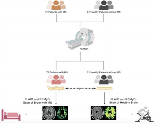
The study by Ouellette et al. (2020) had many parts, the first of which was multiple forms of staining allowing colored dye to reveal the presence and location of myelin within sections of brain tissue examined under a microscope. The next step was recruiting 21 healthy individuals and 71 individuals with MS to participate in the study. As stated earlier, REMyDI combines ideas from many previous studies aimed at measuring myelin, such as quantitative MRI, MTI, T/T relaxometry, and proton density mapping. The simplest way to understand what is being measured by these tests is to understand the nature of water within myelin. The specific properties of water in contact with fatty tissue, as opposed to water in an environment where fatty tissue is not present or is negligible, allows a ratio to be composed, which compares these two situations. This is done using the properties of magnetic fields and the tendency of molecules to spin as they interact with one another, giving a measurable way to assess the state of the myelin present in their brain cells. All MS patients were also given tests by a qualified physician to determine their physical and cognitive disability levels at the time of the study and also approximately 1.5 to 2 years after. The results of the myelin stains and the REMyDI scans were compared, and then quantitatively analyzed in the context of the cognitive and physical disability scores.
The results of the study showed that the myelin found in the staining procedure and in the REMyDI scans had significant correlation in that both methods produced accurate representations of myelin distribution within the brain. Likewise, the disability tests required for the MS patient group aligned with the composition and amount of myelin present at the time of testing and two years after for the follow-up.11 In other words, low myelin amounts in the brain correlated with impaired physical and cognitive ability. The researchers posited that because there appeared to be less myelin in the unlesioned fatty tissue of the brain of MS patients, REMyDI could assess the status of myelin within the tissue, which appeared normal upon cursory visual analysis.
When researching myelin and its relationship to MS, it is inevitable to come across speculation of repairing the damaged myelin as a treatment. The amount of myelin and visible lesions in the brain are also taken as indicators of MS using conventional MRI.7 However, this does not account for the invisible damage to myelin which often accompanies MS. While other studies have explored each aspect of myelin imaging, REMyDI is the first to combine all of these facets into a single comprehensive testing scan which takes 7 minutes and has proven consistent when tested against itself in multiple trials.1,3,5,8 As most scans are expensive and timeconsuming, a single scan will prove useful for future diagnostic and treatment efforts related to myelin diseases. This study was extremely thorough and addressed all previous research done in similar areas while making sure to note differences and explain why they occurred. Comparing multiple myelin stains with actual images from REMyDI, and showing the correlation between them, allows for confidence when addressing the quantities of myelin within specific areas of the brain. The correlation of less myelin with impaired physical and cognitive disability shown by REMyDI and the disability test itself aligns with expectations of MS damaging myelin within the brain, which bodes well for the use of REMyDI in any future studies exploring MS.
[+] References
Ouellette, R., Mangeat, G., Polyak, I., Warntjes, M., Forslin, Y., Bergendal, Å., & … Granberg, T. (2020). Validation of rapid magnetic resonance myelin imaging in multiple sclerosis. Annals of Neurology, 87(5), 710-724.
Vavasour, I. M., Clark, C. M., Li, D. K. B., & MacKay, A. L. (2006). Reproducibility and reliability of MR measurements in white matter: Clinical implications. NeuroImage, 82(2), 637-642.
Warntjes, M., Engström, M., Tisell, A., & Lundberg, P. (2016). Modeling the presence of myelin and edema in the brain based on a multi-parametric quantitative MRI. Frontiers in Neurology, 7(16), 1-15.
Henkelman, R. M., Stanisz, G. J., & Graham, S. J. (2000). Magnetization transfer in MRI: A review. NMR in Biomedicine, 14(2), 57-64.
Hagiwara, A., Hori, M., Kamagata, K., Warntjes, M., Matsuyoshi, D., Nakazawa, M., & … Aoki, S. (2018). Myelin measurement: Comparison between simultaneous tissue relaxometry, magnetization transfer saturation index, and Tw/Tw ratio methods. Scientific Reports, 8(10554), 1-12.
MacKay, A. L. & Laule, C. (2016). Magnetic resonance of myelin water: An in vivo marker for myelin. Brain Plasticity, 2(1), 71-91.
Kolind, S., Matthews, L., Johansen-Berg, H., Leite, M. I., Williams, S. C. R., Deoni, S., & Palace, J. (2012). Myelin water imaging reflects clinical variability in multiple sclerosis. NeuroImage, 60(1), 263-270.
Laule, C., Vavasour, I. M., Moore, G. R. W., Oger, J., Li, D. K. B., Paty, D. W., & MacKay, A. L. (2004). Water content and myelin water fraction in multiple sclerosis: A Trelaxation study. Journal of Neurology, 251(3), 284-293.
Alonso-Ortiz, E., Levesque, I. R., & Pike, G. B. (2015). MRI-based myelin water imaging: A technical review. Magnetic Resonance in Medicine, 73(1), 70-81.
Horsfield, M. A., Barker, G. J., Barkhof, F., Miller, D. H., Thompson, A. J., & Filippi, M. (2003). Guidelines for using quantitative magnetization transfer magnetic resonance imaging for monitoring treatment of multiple sclerosis. Journal of Magnetic Resonance Imaging, 17(4), 389-397.
Sled, J. G. (2017). Modeling and interpretation of magnetization transfer imaging in the brain. NeuroImage,182 (2018), 128-135.
[+] Other Work By Susannah Schloss
Preventing Misdiagnosis of Multiple Sclerosis with CVS (No, Not the Pharmacy)
Neurophysiology
An MRI biomarker was shown to largely prevent misdiagnosis of multiple sclerosis in a real world demonstration of its efficacy.